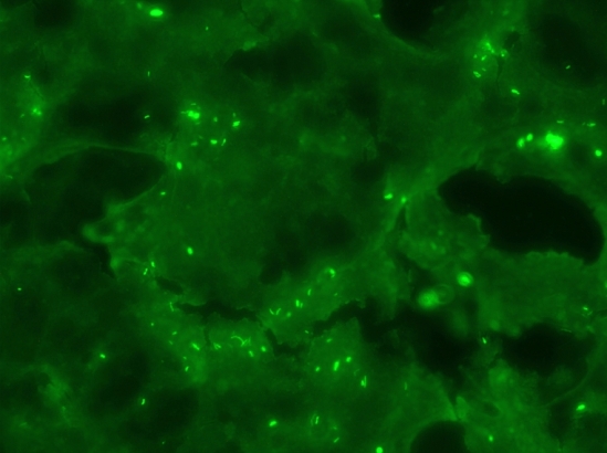
Rabies-positive brain sample. Photo is taken through a fluorescent microscope. The brain sample from the rabid animal was prepared on glass slides, incubated with fluorescent antibodies that attach to the virus, and then viewed through the microscope. The rabies virus is seen as small fluorescent clusters throughout the sample.
Photo Courtesy Los Angeles County Public Health Laboratory, 2007.